Arthritis can severely impact mobility. Many individuals with past sports injuries or those who participate in high-impact activities (like tennis) eventually require joint replacements in later years. When joint replacements occur—whether shoulder, knee, finger joints, or elsewhere—there is substantial mechanical and structural trauma to the body during surgery.

Body Response to Trauma
The body does not distinguish a planned surgical procedure from an accidental injury. It responds to surgical trauma with an acute inflammatory cascade, characterized by the accumulation of fluid and swelling. Ironically, what we often view as a negative side effect is actually a required biological process that builds the physical framework for new tissue. Completely preventing this acute response would stall or entirely halt the healing process.
Why Protein-Rich Fluid is Beneficial and Necessary
When tissue is cut during surgery, blood vessels briefly constrict and then dilate, becoming highly permeable or “leaky.” This allows plasma fluid packed with specialized proteins to flood the interstitial space around the wound, serving critical biological functions:
- Building the Physical Scaffold: This protein-rich fluid contains high concentrations of fibrinogen. Once it leaks into the wounded tissue, it converts into a sticky fibrin mesh. This mesh acts as a literal scaffolding or biological glue that closes the gaps between cut tissues and provides a physical pathway for new cells to crawl across and rebuild.
- Fueling Cellular Growth: The fluid carries proteins, including albumins and globulins, which maintain local osmotic pressure and provide the essential amino acid building blocks that local cells, such as fibroblasts, need to manufacture new collagen.
- Transporting Growth Factors: It functions as a specialized chemical highway. Soluble signaling molecules, cytokines, and growth factors travel through this fluid to signal the regional cellular network exactly how much tissue needs to be repaired.
- Defending the Wound: The swelling creates a localized, high-pressure zone that slows down local circulation just enough to allow white blood cells, such as neutrophils and macrophages, to adhere to the vessel walls and migrate directly into the injury site to destroy invading bacteria.
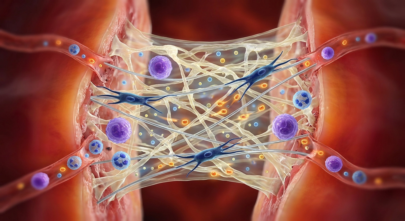
Depiction of swelling and the physical scaffold
Wouldn’t It Be Better to Reduce It?
It is a delicate balance of timing and volume, rather than a question of eliminating the fluid entirely:
- In the First 24–72 Hours: You do not want to completely eliminate inflammation or swelling. Stopping it aggressively during this phase can result in poor wound healing, weak scar tissue formation, or a failure of tissues to knit back together.
- After the First 72 Hours: Once the fibrin scaffold is built and the cellular cleanup crew has finished, the protein-rich fluid has served its purpose. If it stagnates and stays in the tissue too long, it thickens, causing chronic stiffness, severe pain, and a buildup of pressure that cuts off the oxygen supply needed for the next stage of healing.
To prevent this stagnation from occurring, post-surgical rehabilitation relies on a strategic sequence of medical interventions—including early vasoconstriction, targeted compression, anti-inflammatories, and specialized drainage techniques. [image]
These treatments are not designed to stop the natural healing process from happening. Instead, they are used to cap the peak of the swelling so that it does not become so overwhelming that it compresses local capillaries and collapses the very circulation required to deliver these healing proteins.
Manual Lymphatic Drainage (MLD) After Surgery
Manual Lymphatic Drainage (MLD) is a specialized physical intervention that becomes highly beneficial once the initial acute inflammatory influx begins to stabilize, generally around or after the 72-hour mark. Depending on the specific surgical procedure and surgeon clearance, it can sometimes begin as early as 24 to 48 hours post-op to manage rapidly accumulating fluid.
The primary reason why early interventions like ice and compression are used immediately, rather than delayed until after 72 hours, comes down to the physiological difference between fluid prevention versus fluid clearance.
1. The Timeline Shift: Prevention vs. Moving Fluid
Ice and compression are used immediately to control the inflow of fluid, while MLD is used later to manage the outflow.
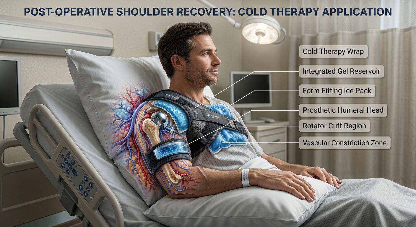
- 0 to 72 Hours (The Inflow Phase): The body is aggressively opening up blood vessels to dump fluid and proteins into the wound. Cold therapy and early compression work right away to narrow these blood vessels and physically squeeze the tissue space. This acts as a barrier that caps the peak amount of fluid that can pool in the first place. If you waited 72 hours to start compression and ice, the tissue would already be massively engorged, and the initial opportunity to limit fluid accumulation would be lost.
- After 72 Hours (The Outflow Phase): Once the fluid is already inside the tissue and the blood vessels have stabilized, cold therapy loses its primary benefit. You cannot freeze fluid out of a tissue. This is where MLD comes in. MLD does not stop fluid from entering; it uses gentle, rhythmic movements to stretch the skin, open damaged or sluggish lymphatic channels, and push the trapped fluid out of the area so the body can reabsorb it into central circulation.
2. The Danger of Ice After 72 Hours
Using ice aggressively after the 72-hour window can actually work against recovery. Once the initial swelling peak passes, the body needs robust blood flow to deliver oxygen and fresh nutrients to repair the cellular matrix. Because ice constricts blood vessels, leaving it on long-term slows down local circulation. This can cause the stagnant, protein-rich fluid to thicken and become gelatinous, which leads to tissue hardness (fibrosis) and severe joint stiffness.
3. The Continuous Role of Compression
Unlike ice, compression is used heavily after 72 hours, often for weeks or months. However, its purpose changes across the timeline:
- Early on (0–72 hours): It provides high resistance to physically stop fluid from pooling in the empty spaces left by surgical dissection.
- Later on (Post-72 hours): It acts as a supportive sleeve that maintains a baseline pressure, preventing fluid from backsliding into the area after MLD or active movement has successfully cleared it out.
Ultimately, medical protocols treat recovery like a construction site: ice and early compression act as floodgates to keep the initial wave manageable, while MLD acts as the drainage pump to clear the site once the storm has passed.
Note: According to 2026 revision of the American Physical Therapy Association (APTA) Clinical Practice Guideline (CPG) for total knee arthroplasty (TKA), published in Physical Therapy (PTJ), it lists updated recommendations post-op TKR. It notes there isn’t sufficient research supporting MLD (among other modalities like Kinesio taping). However, MLD is an adjunct to joint replacement speeding up the body’s natural response,1 & MLD may even be necessary. For example, in a patient who presents with a history of underlying venous stasis, or localized, hyper-inflammatory hard edema. To mitigate the physiological risk of localized lymphostasis, reflex vascular spasm, and subsequent subcutaneous fibrosis as detailed in established lymphology frameworks, manual lymphatic drainage is indicated to restore baseline transport capacity and permit functional progression of range of motion. As the Compendium of Dr. Vodder’s Manual Lymph Drainage states, “In inflammatory processes, protein-rich filtrate gets into precapillary tissue because of histaminic effects. The reflex spasm of the efferent lymph vessels prevents the increased filtrate from being caried off. This can lead to tissue fibrosis, which then impedes the filling of the initial lymph vessels. A further consequence is possible damage to the endothelial cells of the lymph angions. Therefore, after an inflammation has run its course, one often finds fibrous and obliterated lymph vessels in the subcutis cell tissue with diminished or nonexistent functionality. If tissue with a lymphostatic edema becomes inflamed, then lymph transport will become even more severely impaired.”9, 10 The problem is we need more research studies using (proper) MLD post-op to prove the efficacy of MLD.
References
1. Földi M, Földi E, Strössenreuther RHK, Kubik S, eds. Földi’s Textbook of Lymphology: For Physicians and Lymphedema Therapists. 3rd ed. Elsevier Urban & Fischer; 2012. (see pgs. 219-222 Physiological Responses to Impending Lymphostasis)
2. Guo S, Dipietro LA. Factors affecting wound healing. J Dent Res. 2010;89(3):219-229. doi:10.1177/0022034509359125
3. Kumar V, Abbas AK, Aster JC, eds. Robbins & Cotran Pathologic Basis of Disease. 10th ed. Elsevier; 2021:69-105.
4. Szolnoky G, Lakatos B, Keskeny T, et al. Effectiveness of manual lymphatic drainage in intensive phase I therapy of breast cancer–related lymphedema—a retrospective analysis. Breast Cancer. 2024;31(1):122-130. doi:10.1007/s12282-023-01512-y
5. Ebert JR, Joss B, Jardine B, Wood DJ. Randomized trial investigating the efficacy of manual lymphatic drainage to improve early outcomes after total knee arthroplasty. Arch Phys Med Rehabil. 2014;95(9):1743-1750. doi:10.1016/j.apmr.2014.04.024
6. Ultimate Health Clinic. Lymphatic drainage after surgery: what to expect. Ultimate Health Clinic Blog. Published October 21, 2026. Accessed October 26, 2026. ultimatehealthclinic.ca
7. Mechanics of mechanotherapy: post-surgical swelling management with physical therapy. MechanoTherapy Physio. Published March 20, 2026. Accessed October 26, 2026. mechanotherapy.physio
8. Bove AM, Carroll LA, Cone S, et al. Clinical practice guideline for physical therapist management of total knee arthroplasty: revision 2026. Phys Ther. 2026;106(7):pzag058. doi:10.1093/ptj/pzag058
9. Kasseroller R. Compendium of Dr. Vodder’s Manual Lymph Drainage. Karl F. Haug Verlag; 1989. (see page 89)
10. Kurz, I.: Lehrbuch der Manuellen Lymphdrainage nach Dr. Vodder, BD. 2 und 3 [Textbook of Dr. Vodder’s Manual Lymph Drainage, Vols. 2 and 3]. Karl F. Haug Verlag, Heidelberg 1989.