Jackie’s Story (part 3)
Jackie’s Story (part 2)
This is Part 2 of the first person I ever knew with lymphedema: my high school English teacher (Jackie Miller). Doctors weren’t sure why her leg was swelling. Was it cancer recurrence? A blood clot? An ovarian cyst? Scar tissue? Or possibly something else? (Jackie was referred to as Janie’s earlier in the blog for anonymity. With the family’s permission, her real name is being used.)
It’s here!
Jackie’s Story (part I)
November was the first blog I’ve missed in a long time! Figuring out how to compile and edit this video in a story format was more work than I anticipated. But part one is finished. Take a moment to watch, “like,” and subscribe.
(Jackie was referred to as Janie’s earlier in the blog for anonymity. With the family’s permission, her real name is being used.)
Jackie’s Story (video pending)
Jackie’s story of cancer & lymphedema coming in December. (Jackie was referred to as Janie’s earlier in the blog for anonymity. With the family’s permission, her real name is now being used.)
November will not have a post. Learning video editing is more arduous than expected! Stay tuned…
Janie’s Story (Part 2)
In September, we talked about my high school English teacher, Janie.* She had developed lymphedema due to her cancer but also had other contributing factors to her lymphedema development. Let’s continue with additional contributing factors to her lymphedema development. We’ll also see how she managed her swelling over the years. Read Part 1 here.
Diuretic Use
Lasix was prescribed as a treatment for Janie’s leg swelling most likely in 1982. Lasix is a diuretic used to get rid of excess fluid. Diuretics are a contraindication for the treatment of lymphedema when that is the sole condition being treated. 3 The reason is while diuretics do help get rid of excess fluid by increasing urine output, they cause hemoconcentration (an increase in the concentration of what’s in blood other than fluid – like the protein mentioned previously). The lymphatic system transports several items from the tissue. One of those items happens to be protein.
Protein attracts water. If the lymphatic vessels of an extremity are damaged and proteins are accumulating in the tissue, what happens? Fluid begins to accumulate (swelling). The definition of lymphedema is an accumulation of protein-rich fluid caused by a structurally impaired lymphatic system. As protein, fluid & other matter that the lymphatic system normally transports remain in the tissue, inflammation develops. This leads scarring or fibrosis (hardening) as the body remodels the tissue. This process causes the tissue to become firmer over time & creates more congestion.
Cancer return
In 1984, Janie admitted to the hospital with shortness of breath & chest pain.
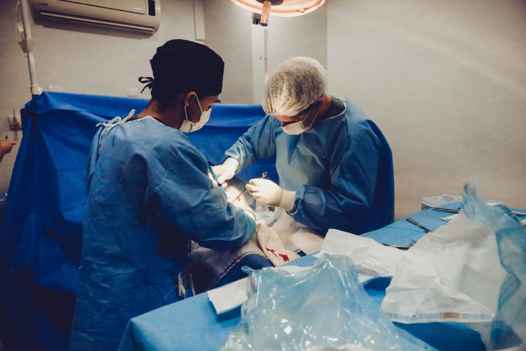
She was found to have a pleural effusion (a buildup of fluid between the lung(s) & the outer lining), atelectasis (partial lung collapse), pericardial effusion (fluid around heart), enlarged chest lymph nodes and a mass in her chest. As a result of the fluid around her heart, she developed pericardial tamponade (a condition which prevents the heart from fully refilling). Blood pressure can drastically drop & cause death. She had an emergency procedure in which the surgeon cut into her chest between the ribs to access the heart. Once there, a cut was made into the lining of the heart to drain the excess fluid. A biopsy of the mass was done & chest tubes were inserted to allow draining after surgery. The biopsy revealed lymphoblastic lymphoma. Six months of chemo, radiation to her head & several spinal chemo injections later, Janie recovered & remained in cancer remission for several years.
Lymphedema
Non-pitting, chronic lymphatic obstruction
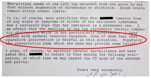
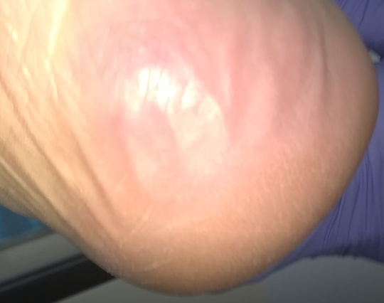
Medical records (below) describe the swelling in her left ankle as not being painful. Other than the early stage of congestion, lymphedema is generally not painful. (An exception may be when there is edema & lymphedema.) The spongy pitting becomes firmer over time until an indentation can no longer be made, & eventually, swelling no longer reduces with elevation. In 1987, a doctor’s letter describes Janie’s lymphedema as non-pitting, extending from the groin to the foot, lymphatic-obstruction, and not reversible. She is prescribed a pump.
Intermittent Pneumatic Pump
Dennis (Janie’s husband) described this pump as one that encompassed her leg only (no trunk component) & started pumping from the foot & continued up the leg. This is a typical sequential that can be helpful for venous insufficiency, but it isn’t the right pump for lymphedema. In fact, it can worsen lymphedema instead of improve it.4 Here’s why:
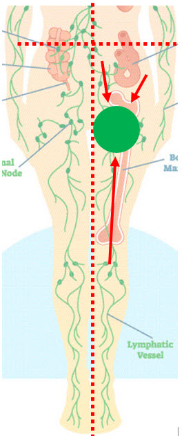
In cases of lymphedema, a quadrant of the trunk is usually involved in addition to the swollen extremity.
Both the quadrant & extremity drain to the same lymph node region. In Janie’s case, her left leg & left lower quadrant of her trunk would have been involved. These areas drain to the same area (the left inguinal lymph nodes). If a pump leg sleeve stops near the thigh, fluid is just pushed the drainage region that is already impaired. Janie’s pump was prescribed in the 1980s, but lymphedema patients today are often prescribed a similar pump.
While the pump may decongest the leg temporarily, it ends up pushing fluid (& shifting pressure) to the already congested area of trunk quadrant & inguinal lymph nodes. In cases of leg lymphedema, fluid may even be shifted into the genitals. If it backs up into the trunk quadrants, a fibrosclerotic wall develops. Foldi’s Textbook of Lymphology outlines additional concerns: worsening lymphedema, removing more water than protein & increased pulmonary blood capillary pressure.5
Recommended pumps will have an abdominal trunk component & initiate a proximal massage before pumping. Ideally, these pumps will decongest the trunk, upper thigh, lower, thigh, knee, upper lower leg & lower leg (in that order) before beginning to push fluid from the foot up. This “sort of” mimics the manual lymph drainage sequence & decongests the area before pushing fluid into it from the swollen leg.
Compression Garment
In 1997, a physician wrote a letter that summarized Janie’s status. They noted that she wore TED hose & used ACE bandages. If she really was using ACE bandaging, she would see some improvement but not as good as she would with traditional multi-layer, short-stretch lymphedema compression. ACE wraps are long-stretch bandages which have more elasticity & give under resistance. They don’t provide the working pressure short-stretch bandages do to remove fluid from the tissue as a person uses their muscles. Some patients also do not tolerate long-stretch as well.

The compression hose she used per this note were TED hose.
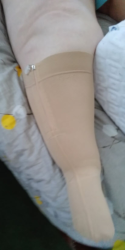
TED hose are anti-embolism hose often used in hospitals after surgery. They have 15-20 mmHg which isn’t typically enough compression for cases of leg lymphedema. Generally, if there is no arterial blood flow impairment or other concerns, compression of 30-40+ mmHg are used on a leg to overcome pressure in the tissue caused by gravity. I know Janie never obtained a custom, flat-knit hose when I was in school (the recommended garment). She used a circular-knit hose which looks similar to a pantyhose & don’t provide the same benefits that custom, flat-knit hose do.
Lymphedema Treatment
Janie did attend a lymphedema clinic in St. Louis in the late 1990s / early 2000s (which was an hour & a half away from her home – the nearest one). Lymphedema treatment includes a massage called manual lymph drainage (MLD), multi-layer short-stretch bandages, exercise & skin care. Records reveal insurance began paying for MLD in the late 90s. I’m hopeful she may have eventually gotten better care for her lymphedema.
Breast cancer, traffic, blood clot & pelvic cancer
In 1997, she noticed a small nodule in her right breast, but doctors were not concerned. They wanted to monitor it. However, between June of 1999 & July 2000, she was diagnosed with ductal carcinoma in situ (breast cancer). She had a lumpectomy & was prescribed Tamoxifen which appeared to resolve her breast cancer. In 2002, she was stuck in traffic for three hours & began having leg pain. She went to the emergency room & was found to have a DVT (blood clot) in her left leg with significant increased swelling.
She was evaluated by Mayo Clinic for her lymphedema soon after. The doctor stopped lymphedema massage (due to the DVT) but had her continue compression on her leg. According to notes, she never used night compression as a part of her maintenance routine. (Day & separate night compression should be a part of lymphedema maintenance. Without night compression, the leg often refills.)
In 2003, Janie was diagnosed with pelvic cancer. In 2004, she decided to retire a year early & spend time with her family & enjoy traveling (to Ireland, Turkey, the Pacific Northwest…). She saw her son get married, & she met her first grandbaby – no doubt two of the biggest joys of her life. In 2006, the pelvic mass was described as encompassing her common iliac blood vessels & ureter. She received radiation to her pelvis with some improvement (including some leg swelling improvement likely due to tumor shrinkage). In 2009, Janie lost her battle with cancer.
Continued to live a full life
From the beginning of her first cancer diagnosis, Janie kept excellent records allowing me to follow her journey through cancer, treatment, recurrence & complications. Even though she didn’t dwell on her problems, her notes gave me insight into what she was might have been thinking at times as she oriented to the medical terms with self-education. Titles of some of her printouts included:
- side effects of chemo: hair loss, low WBC count, neuropathy, joint pain, muscle pain, cardiac effects, local irritation, low RBC count, stomach upset, diarrhea
- information about metastatic cancer
I can only imagine the self-consciousness a person experiences losing their hair let alone the fear that can come from a diagnosis of cancer. No doubt she struggled with the diagnoses she faced. She had several setbacks besides those already mentioned. Over the years she also experienced chronic anemia; short-term facial swelling; acute Bell’s Palsy; enlarged spleen; mild, chronic left hip pain from bursitis (& likely from the weight of her swollen leg); a blocked ureter (impeding urine flow from one kidney to the bladder); a failed ureter stent placement; urinary tract infection(s); shingles & a severe pelvic infection which became septic — & that isn’t all. I had no idea what she went through. She dealt with these challenges with strength & dignity. Never once do I recall her complaining. Dennis said she wasn’t one to focus on herself or her problems. Instead, she chose to face the obstacle, seek resolution or management, do what was best for her family & focus on living.
This series is dedicated to “Janie” & all those who are managing lymphedema caused by cancer.
~Janie~
(11/01/50 – 3/12/09)
I’ll be featuring my interview with Dennis in the coming YouTube channel. Stay up-to-date on development by joining the conversation.
References
1 Bankov K, Döring C, Ustaszewski A, Giefing M, Herling M, Cencioni C, Spallotta F, Gaetano C, Küppers R, Hansmann ML, Hartmann S. Fibroblasts in Nodular Sclerosing Classical Hodgkin Lymphoma Are Defined by a Specific Phenotype and Protect Tumor Cells from Brentuximab-Vedotin Induced Injury. Cancers (Basel). 2019 Oct 30;11(11):1687. doi: 10.3390/cancers11111687. PMID: 31671543; PMCID: PMC6896072. Available from: https://pubmed.ncbi.nlm.nih.gov/31671543/.
2 Kamal MM, Khude SR, Yadav SB, Raut WK, Pangarkar MA. Syncytial variant of nodular sclerosing Hodgkin’s disease: A diagnostic pitfall in fine-needle aspiration cytology. J Cytol. 2014;31(2):91-92. doi:10.4103/0970-9371.138674. Available from: https://www.ncbi.nlm.nih.gov/pmc/articles/PMC4159904/.
3 Foldi, M, Foldi, E. (2006). Foldi’s Textbook of Lymphology (2nd ed.), p. 282-283, 446. Germany: Urban and Fisher.
4 Foldi, M, Foldi, E. (2006). Foldi’s Textbook of Lymphology (2nd ed.), p. 282. Germany: Urban and Fisher.
5 Aldrich MB, Gross D, Morrow JR, Fife CE, Rasmussen JC. Effect of pneumatic compression therapy on lymph movement in lymphedema-affected extremities, as assessed by near-infrared fluorescence lymphatic imaging. J Innov Opt Health Sci. 2017;10(2):1650049. doi:10.1142/S1793545816500498
Janie’s Story (Part 1)
Janie* was my high school English teacher. She is also the first person I knew who had lymphedema (though I didn’t know it at the time). I recall seeing her walk down a hallway at school & noticing her left leg was larger than the right (she often wore skirts and dresses). I wondered why but never really gave it much thought. She was just “Mrs. Mitzer,” my English teacher. Another occasion, I noticed she wore a hose on her left leg that was stretched to the point that tiny gaps in the fabric could be seen. It would be years before I would understand why Janie’s leg was larger & why her sock was so stretched.

A photo of Janie from my high school year book
Not long ago I decided to transition in business with the purpose of raising awareness about lymphedema & being able to help a greater number of people. One way I am doing that is by telling the stories of people who have lymphedema & developing educational videos. Many of these stories will be from people I have treated. But this story is different. Given my new venture, it seemed fitting to pay tribute to the past & recognize Janie since she was the first person I knew who had lymphedema. Regretfully, she passed away in 2009. I wondered if her family would be willing to talk about her experience with lymphedema. I contacted her husband, Dennis, earlier this year.
The interview
Dennis graciously obliged. He has since remarried to Jane. Both Jane & Dennis were present for the discussion. The first question Dennis asked me when he saw my notepad was, “Is that notepad filled with questions?” I assured him it wasn’t. The notepad did have a few questions, but it was mostly filled with notes I had taken while reviewing Janie’s medical records which he had provided me before our interview.

The beginning
Janie’s medical story begins in 1979 when she was diagnosed with stage 4B, nodular sclerosing classical Hodgkin lymphoma. At the time Janie’s cancer was found, it had spread beyond her lymph nodes & throughout her abdomen. Dennis remembered Janie waking up at night sweating, her pillow & night clothes soaked. Indications of Hodgkin lymphoma can include night sweats, fever, weight loss & other symptoms. She received several chemotherapy treatments & eliminated all signs of cancer (as a 1980 exploratory laparotomy confirmed). One of the potential consequences of Hodgkin lymphoma is secondary cancer including, but not limited to, breast cancer. That would later prove true in Janie’s case.
First signs of swelling
In April of 1982, Janie noticed swelling in her left ankle, but it resolved with elevation. Medical records indicate the swelling was graded at 2+. This means a finger could be depressed into the skin slightly (less than ¼ of an inch) or the tissue would hold a depression for up to a max of 15 seconds. There was fullness in the left lower part of her abdomen as well. The initial fear was lymphoma recurrence, but a CT scan & exploratory surgery of her left groin (inguinal lymph nodes) ruled this out. Imaging did reveal an enlarged left ovarian cyst. Doctors felt her leg swelling might be due to venous obstruction caused by this cyst putting pressure on her [left iliac vein] or due to scar tissue.
However, a medical note I read indicated suspicion of a superficial blood clot. A superficial clot is usually less concerning than a deeper one (called a deep vein thrombosis or DVT). Symptoms of a blood clot generally include swelling as well as pain, redness & warmth. Janie’s only symptom was swelling which initially went down with elevation. A venogram did not show a definitive clot, but this theory persisted in her medical records.

Suspect superficial thrombophlebitis
The name of the cancer Janie had, “nodular sclerosing,” indicates its behavior.1, 2 This cancer creates fibrosis and sclerosis or thick bands of collagen (scar tissue) in the lymph nodes. This would likely contribute to lymphedema in her left leg as it would likely impair lymph node function.
Other contributing factors
Janie also had several surgeries in the left inguinal & pelvic areas over time which would cause trauma & scarring. Between 1982 & 1984, she developed an infection in her left leg (twice) called cellulitis. She was a teacher & spent a good deal of time standing and sitting, increasing the pressure in her legs. Veins in the legs must overcome the pressure caused by gravity in order to return fluid to the heart. Over time, those veins begin to tire & weaken. The lymphatic system takes up the slack for a while, but just like veins, they can fatigue & weaken also. When they do, scarring ensues as molecules they normally remove now remain in the tissue. All these scenarios could have caused her lymphedema to get worse.
To be continued 10/01/21.
References
1 Bankov K, Döring C, Ustaszewski A, Giefing M, Herling M, Cencioni C, Spallotta F, Gaetano C, Küppers R, Hansmann ML, Hartmann S. Fibroblasts in Nodular Sclerosing Classical Hodgkin Lymphoma Are Defined by a Specific Phenotype and Protect Tumor Cells from Brentuximab-Vedotin Induced Injury. Cancers (Basel). 2019 Oct 30;11(11):1687. doi: 10.3390/cancers11111687. PMID: 31671543; PMCID: PMC6896072. Available from: https://pubmed.ncbi.nlm.nih.gov/31671543/.
2 Kamal MM, Khude SR, Yadav SB, Raut WK, Pangarkar MA. Syncytial variant of nodular sclerosing Hodgkin’s disease: A diagnostic pitfall in fine-needle aspiration cytology. J Cytol. 2014;31(2):91-92. doi:10.4103/0970-9371.138674. Available from: https://www.ncbi.nlm.nih.gov/pmc/articles/PMC4159904/.
(Part 4) You’re Just Fat…or Are You?
Read Part 3 here
Continuing lymphedema treatment
At the conclusion of July’s post, Carol was waiting for her hose. They arrived & she loved them! One problem: The manufacturer didn’t sew the ankle pocket around the full ankle as requested. Turns out, this manufacturer couldn’t sew a pocket all the way around the ankle. Why didn’t they mention that before? Without the pocket, the hose would slip down & bunch, causing pain.
Long story short, I complained, sending copies of communications with the manufacturer’s customs department describing the need for a full pocket and why. Their response was fantastic. They gave us a refund due to the situation. Carol and I tried two more manufacturers and finally got what she needed. We’re still working on adjustments, but she’s very happy with the containment and feel of the compression garments.
The hose were one of several impediments Carol has faced. I mentioned some of them in July. Let’s look at a few more.
Complication – diarrhea
Carol went back to a Keto diet she had used to lose weight in the past (the obesity weight, not the lipedema weight). The diet calls for ingesting a particular oil and liquids the first week which gave her diarrhea. Sometimes that diarrhea ended up on leg bandages. So, she would remove the bandages to shower and her legs would refill. Eventually, she progressed in her diet and recovered from that complication.
Complication – emotional upheaval
Losing someone close to her earlier this year, she has struggled with the void their passing created. In addition, she had a major life change – her job. Covid caused a lot of people to feel isolated as they worked from home. Carol felt isolated, too. After months of structural change (which included moving people she had worked with for years to other departments), she decided to retire. Now she was not only home and alone much of the time, she also wasn’t interacting with the people she enjoyed talking to. Carol is an extrovert, so this was a hard adjustment. She’s currently pursuing virtual counseling options.
Complication – transient ischemic attack (TIA)
As if the prior complications weren’t enough, she spent a weekend in the hospital after experiencing symptoms of a stroke, including spiked blood pressure and difficulty talking (in this case, the speech difficulty was expressive aphasia). She had just been to see her doctor a few weeks before. She was told her cholesterol was elevated and she was given a new medication to help. Then she awoke one Saturday morning unable to talk.
She was taken to the ER by ambulance. After a day of monitoring, they released her with inconclusive results that pointed to a TIA. A TIA is often referred to as a “mini-stroke.” Symptoms are less severe than a stroke and don’t usually last long because the blockage in the brain is temporary. In the days that followed, Carol had occasional difficulty pronouncing words and remembering lyrics to some of her favorite songs, but words were coming back to her. Over the next several days, as we worked on her aphasia and as she practiced on her own, speech improved. But not for long.
More recently, she has been complaining of numbness in her face that comes and goes. I noticed her speech seemed to be getting worse in our last session (even when she slowed down). Her doctor wanted her to hold therapy until she consults a neurologist. That is where we are now. Waiting for her hose revisions and waiting to find out what’s causing her aphasia and numbness.
Conclusion of the lipedema blog series
Carol has had several battles during her lymphedema treatment. But her persistence paid off & she’s made some awesome functional gains as she’s gotten her legs under control. She can get up and down from her chair, in and out of bed, and in and out of her car. She drove a couple times (which she hasn’t done in a year). She has more work to do (much of which is on hold pending her neurology follow up), but she’s still motivated — & she’s happy with her lower leg status. I hope to give you a positive update on her condition in the future.
For more information about lipedema, visit the following links: Dr. Karen Herbst, Lipedema Foundation, The Lipedema Project, the Fat Disorders Resource Society, National Center for Advancing Translational Sciences and this article in the Plastics and Reconstructive Surgery Journal.
(Part 3) You’re Just Fat…or Are You?
Read Part 2 here
Getting Treatment
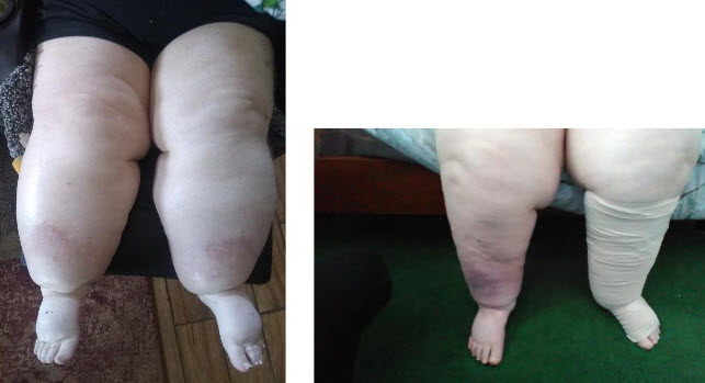
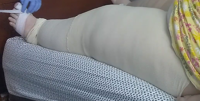
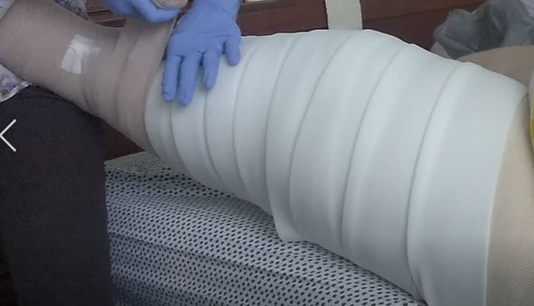
Carol started treatment to reduce her leg size. We began with manual lymph drainage (MLD), multi-layer compression bandaging (using a liner, toe gauze, foam, and short-stretch bandages) three days a week. We added exercise and skin care. (This four-part treatment is called Complete Decongestive Therapy or CDT).
She lost volume quickly – very quickly. In fact, her compression would slip down prior to the next visit allowing refilling. (This was later resolved using an adhesive lotion and an elastic Tubular sleeve.) But progress wasn’t without its complications.
Complication – mobility
Although I recommended Carol start with one leg (bandaging to her groin), she wanted to start with both legs. To avoid returning too much fluid to her heart and kidneys too fast, we bandaged both legs to the knee only the first week of treatment. Bandaging both legs made getting up from her chair more difficult because it limited ankle flexion. Carol later decided she wanted to bandage one leg only. As a result of straining to get up from her chair, she developed biceps tendonitis, causing shoulder pain.
Complication – skin integrity
Carol also found getting to the bathroom promptly was a challenge (causing her to soil herself and the thigh-high bandages). Because she hadn’t performed her normal hygiene routine, she developed a yeast infection and skin breakdown under her abdominal fold.
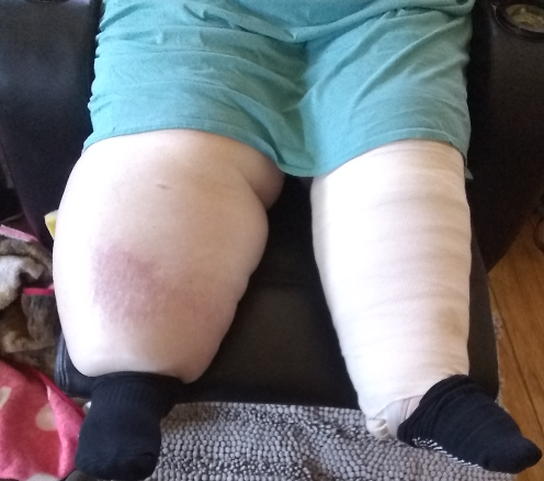
Complication: refilling and weight gain
Unhappy with her hygiene status, Carol began removing her compression to shower before I arrived, allowing her leg to refill. Despite encouragement to continue thigh-high bandaging with hygiene modification (such as using a cast protector to shower), she decided she wanted to regress bandaging to the knee only. As a result, her thigh refilled and she regained some of the weight she had lost. Because of the atypical leg shape, bandaging was tricky & a few times she had heel pain due to pressure. This problem was resolved by fabricating a doughnut foam shape and placing it under her compression, offloading the pressure to her heel.
Complication – shoulder pain
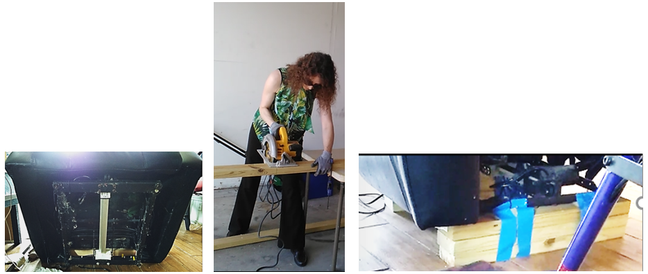
Carol’s shoulder pain persisted. We raised the height of her chair by adding pillows, but she couldn’t get comfortable due to hip arthritis pain. I encouraged her to modify her chair height by letting me install 2x4s. She declined feeling this was regression instead of progress. (She felt she just needed to be stronger.) Several sessions later, as her shoulder pain got worse and as she became frustrated having to ask her spouse for help getting up, she agreed to modify her chair. She later described this modification as “life changing.” When she became compliant applying ice to her shoulder to reduce inflammation, and as she continued to use the 2x4s installed for chair height elevation, her shoulder pain went away.
We added exercise to her bandaging, and she slowly began to see more functional progress. She is now able to get into her bed & into her car on the driver’s side without assistance. While therapy has encountered other complications, at this point, we are waiting for her knee high compression hose (and I’m hoping she will later get custom, flat-knit biker shorts (capri length) with a zipper for full leg & abdominal compression). Stay tuned!
For more information about lipedema, visit the following links: Dr. Karen Herbst, Lipedema Foundation, The Lipedema Project, the Fat Disorders Resource Society, National Center for Advancing Translational Sciences and this article in the Plastics and Reconstructive Surgery Journal.
Blog series to be concluded August, 2021.
- « Previous Page
- 1
- …
- 4
- 5
- 6
- 7
- 8
- 9
- Next Page »