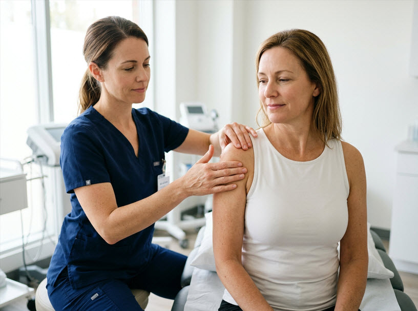
Last month, we started a discussion about MLD pressure. Foldi always taught light pressure. Newer research indicates lymph vessels aren’t occluded until 86 mmHg. We’re continuing that topic this month.
Why This Does Not Mean “Deeper Pressure Is Better”
One of the most important conclusions from the study is often overlooked:
The researchers specifically noted that manual lymph drainage pressures applied by the hand are typically under 10 mm Hg, which remains well below lymphatic occlusion thresholds. (Sage Journals)
That matters because MLD is not intended to “force fluid through pipes.” Instead, it works by:
- stimulating lymphatic vessel activity,
- improving lymphangion filling,
- enhancing rerouting through collateral pathways,
- and supporting natural lymphatic pumping.
The study supports the idea that properly performed gentle MLD does not collapse lymphatics. In other words, Foldi’s gentle-pressure philosophy remains physiologically sound.
The Difference Between Compression and MLD
The study also helps clarify confusion between:
- manual lymph drainage pressure, and
- compression therapy pressure.
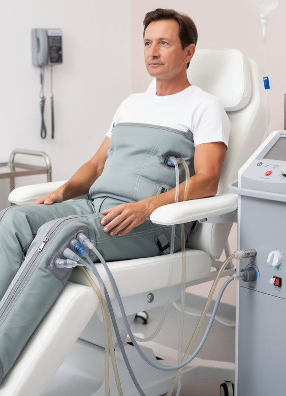
Compression garments and pneumatic compression devices may use substantially higher pressures than hands-on MLD. The researchers warned that if compression pressures exceed lymphatic occlusion pressure, collectors and initial lymphatics could temporarily collapse during inflation phases. (Sage Journals)
This distinction is critical in clinical practice.
Foldi-based Complete Decongestive Therapy (CDT) has always emphasized that:
- MLD prepares and stimulates lymphatic pathways,
- compression maintains fluid reduction,
- exercise activates the muscle and lymphatic pumps,
- and skin care protects vulnerable tissues.
The therapies work together — but they do not work the same way.
What This Means for Therapists Today
The 2016 findings encourage a more evidence-based discussion around lymphatic pressure without abandoning traditional lymphology principles.
For certified lymphedema therapists, the takeaway is not to use heavier pressure. Instead, the study reinforces several clinical ideas:
1. Gentle Pressure Can Still Be Effective – MLD does not require deep force to stimulate lymphatic transport.
2. Vessel Filling Matters – Lymphatic flow is intermittent. Proper sequencing and preparation techniques — central clearing, proximal-to-distal preparation, and rhythmic repetition — may improve lymphatic filling and flow.
3. Precision Matters More Than Intensity – Direction, sequencing, stretch, and rhythm likely influence outcomes more than raw pressure.
4. Compression Must Be Individualized – Higher pressure is not automatically better, especially in compromised or fibrotic tissues.
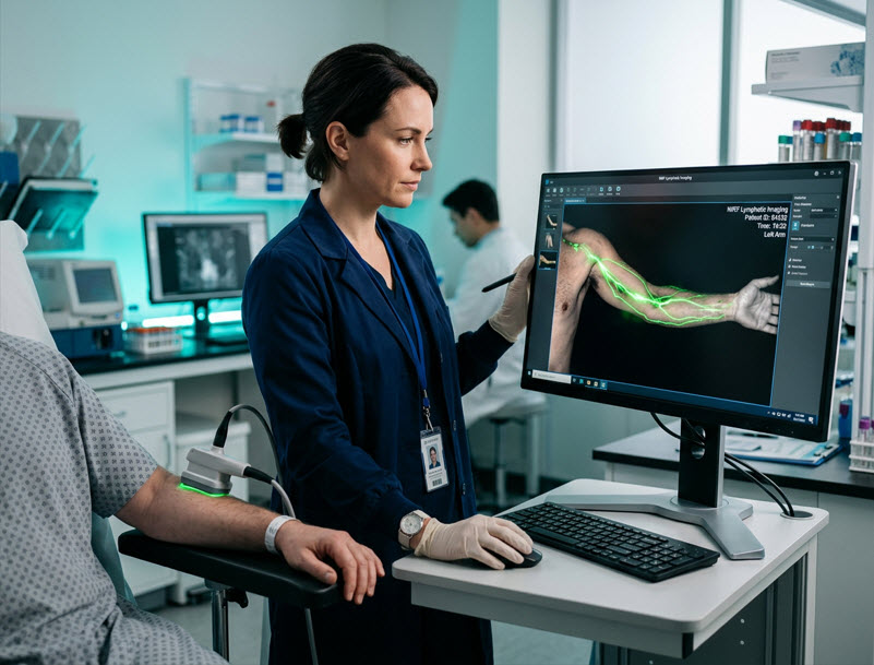
Modern Imaging Is Validating Traditional Lymphology
One of the most fascinating aspects of this research is how modern imaging technology is beginning to visually confirm concepts long taught in Foldi-based education.
The lymphatic system is dynamic, contractile, and responsive — not passive.
Near-infrared fluorescence imaging allows researchers to watch lymphangions contract and observe fluid movement in real time, offering a new scientific window into therapies that were previously understood mostly through clinical outcomes and practitioner experience. (Sage Journals)
As lymphatic science evolves, the future of lymphedema care will likely combine:
- traditional clinical wisdom,
- advanced imaging,
- individualized pressure strategies,
- and deeper understanding of lymphatic biomechanics.
The Foldi principles are not being replaced. They are being explained more clearly than ever before.
References
1 Foldi, M, Foldi, E. (2006). Foldi’s Textbook of Lymphology (2nd ed.), p. 526. Germany: Urban and Fisher.
2 Belgrado, J.-P., Vandermeeren, L., Vankerckhove, S., Valsamis, J.-B., Malloizel-Delaunay, J., Moraine, J.-J., & Liebens, F. (2016). Near-infrared fluorescence lymphatic imaging to reconsider occlusion pressure of superficial lymphatic collectors in upper extremities of healthy volunteers. Lymphatic Research and Biology, 14(2), 70–77. https://doi.org/10.1089/lrb.2015.0040